Salivary Cysts in Dogs
 We frequently see
canine patients at our clinic with a huge, fluid-filled cyst located either
under their jaw or within their mouth. When we say huge, we really mean what we
say because some of those found below the lower jaw are larger than
grapefruits. There are many other causes of swelling in this area such as
puncture wounds, abscesses, infections of the teeth, and tumors, but most
common relate to the salivary glands.
We frequently see
canine patients at our clinic with a huge, fluid-filled cyst located either
under their jaw or within their mouth. When we say huge, we really mean what we
say because some of those found below the lower jaw are larger than
grapefruits. There are many other causes of swelling in this area such as
puncture wounds, abscesses, infections of the teeth, and tumors, but most
common relate to the salivary glands.
The salivary glands, as their name implies, are responsible for the production of saliva. Several sets of them lie in the tissue area beneath the skin in the upper and lower jaw. Saliva production goes on constantly. It continuously makes its way into the mouth through small ducts that originate within the salivary glands and course under the skin, opening directly into the oral cavity. The purpose of saliva is to lubricate the mouth, moisten the food, and begin the digestive process. Saliva breaks down starch into individual sugar molecules, both within the mouth and during chewing, and later on in the stomach and small intestine.
What causes salivary cysts?
Salivary cysts occur when saliva leaks from the salivary ducts into the tissues near or within the mouth. This may be caused simply by trauma to the ducts. In some instances, the duct may have been broken or it may have been bruised with the resultant swelling closing off the duct. In other cases, an abscess or tumor in the area may put enough outside pressure on the duct to pinch it off as you might a garden hose. Finally, there are instances when the glands, because of an internal inflammatory process, release a fluid that is just too thick to make its way through the tiny ducts. This again leads to an obstruction. All of these cases have the same final result. Whether the duct was originally ruptured or not, the pressure of continued saliva production by the glands will finally cause the walls of these tiny tubes to break and allow saliva to leak into the surrounding tissue.
A small amount of a fluid leaking into tissue rarely, if ever, causes a problem and will usually go unnoticed. We often give large quantities of fluids directly under the skin to a dehydrated puppy or kitten. (It is sometimes impossible to find a vein in these tiny animals for the preferred intravenous route.) It is absorbed by the blood system in a few hours and distributed throughout the body. However, saliva leaking into the tissues almost always develops into a major problem because the fluid is produced constantly and in large quantities. Additionally, possibly because of the viscosity, the body has a difficult time reabsorbing it. It simply continues to build and forms large cysts.
What do salivary cysts look like?
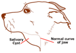
In an affected animal, we see the salivary cysts in either of two places. Typically, a large pocket of saliva will form under the jaw at the base of the neck. It will initially be on the same side as the injured duct, but after a brief period, the cyst can become so large that it fills the entire area below the jaw. It feels like a thick-walled balloon filled with honey. The animal is able to breathe, eat, and drink in a normal fashion. Swallowing rarely seems to be impaired. The other area where cysts are found is within the mouth, under the tongue. They are referred to as a 'ranula' in this location. These cysts often will cause problems with eating and drinking. They may limit normal movement of the tongue during drinking or get in the way when food is being chewed.
What is the treatment for salivary cysts?
When dealing with a salivary cyst, medical therapy alone is rarely successful. There are isolated instances in which a local infection or inflammation can be eliminated with medication. If the salivary duct flow has only been restricted, all may return to normal. Once the salivary duct has ruptured, however, the leakage will continue. We do not know of any case in which a duct closed or healed on its own after injury. The problem does not 'just go away.'
 Surgery is required
in cases where saliva is leaking into the surrounding tissues. In those animals
with a large, fluid-filled cyst below the jaw, the salivary glands on the affected
side are completely removed. Usually this means all of the mandibular and
sublingual glands on one side. Although this may seem extreme, the remaining
glands respond to the increased need and adjust their production to compensate
for the removal. First, the cyst is opened, and the fluid is drained; then
excess tissue is removed and the skin is closed. A second incision is then made
over the main body of glands and they are removed along with a portion of the
duct. This is easier said than done because of the many nerves and major blood
vessels in this area. In cases involving a ranula, the surgery is much more
simple. The ranula is opened and its walls are basically turned inside out and
sutured down to the lining of the mouth. This creates an everted pouch that
slowly heals, forming a new opening for the salivary duct. The salivary glands
themselves are not affected in the surgical correction of a ranula.
Surgery is required
in cases where saliva is leaking into the surrounding tissues. In those animals
with a large, fluid-filled cyst below the jaw, the salivary glands on the affected
side are completely removed. Usually this means all of the mandibular and
sublingual glands on one side. Although this may seem extreme, the remaining
glands respond to the increased need and adjust their production to compensate
for the removal. First, the cyst is opened, and the fluid is drained; then
excess tissue is removed and the skin is closed. A second incision is then made
over the main body of glands and they are removed along with a portion of the
duct. This is easier said than done because of the many nerves and major blood
vessels in this area. In cases involving a ranula, the surgery is much more
simple. The ranula is opened and its walls are basically turned inside out and
sutured down to the lining of the mouth. This creates an everted pouch that
slowly heals, forming a new opening for the salivary duct. The salivary glands
themselves are not affected in the surgical correction of a ranula.
The important point for owners to recognize is that salivary cysts should be seen by a veterinarian as soon as possible. If the owner waits too long in a case of the cyst below the jaw, it is difficult for the clinician to determine on which side it originated. No one wants to surgically remove all the mandibular and sublingual glands from one side and then discover it was the wrong side. Help your veterinarian and your dog with an early trip to the clinic.